ヘリコバクター ピロリ抗体試薬 -一次抗体
陽性対照:
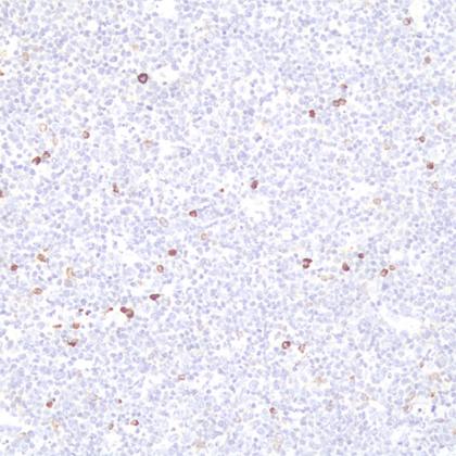
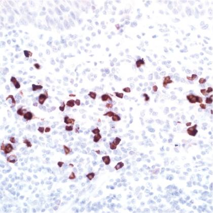
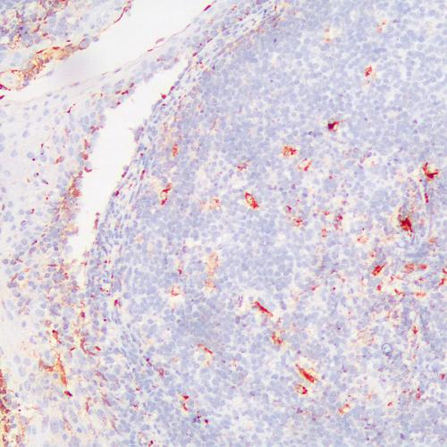
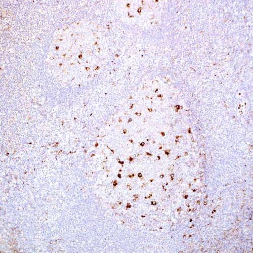
tissue infected by H. Pylori細胞局在化:
H. Pylori応用:
IHC-P二次抗体:
iVision™仕様/ml:
1、3、6、0.2(Concentrated)【Product name】
Helicobacter Pylori Antibody reagent--Primary Antibody
【Packing specification】
| Code | Clone | Specifications |
| AP0133 |
polyclonal |
0.1ml, 0.2ml, 1ml, 1.5ml, 3ml, 6ml, 11ml, 30ml |
| AR0133 |
TLR0133 |
0.1ml, 0.2ml, 1ml, 1.5ml, 3ml, 6ml, 11ml, 30ml |
【Intended use】
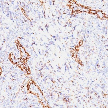
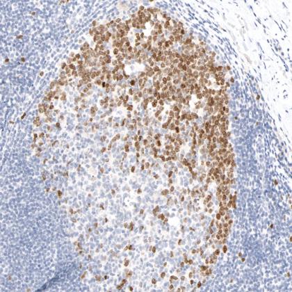
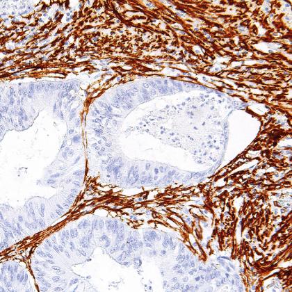
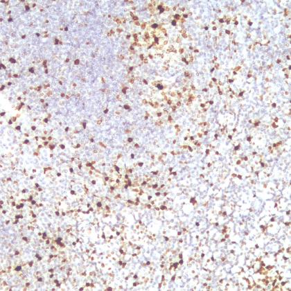
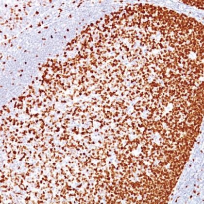
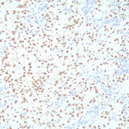
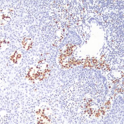
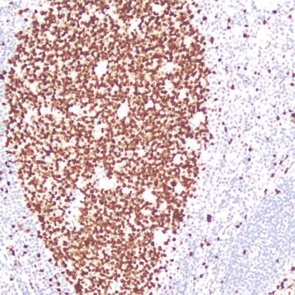
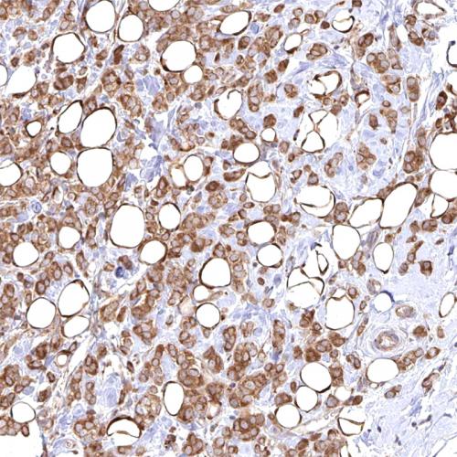
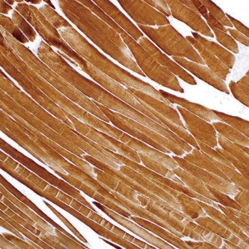
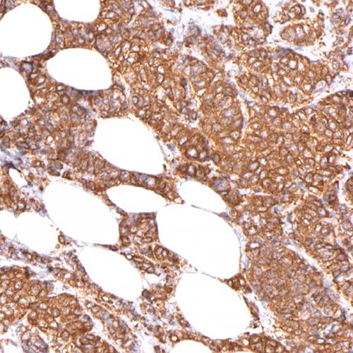
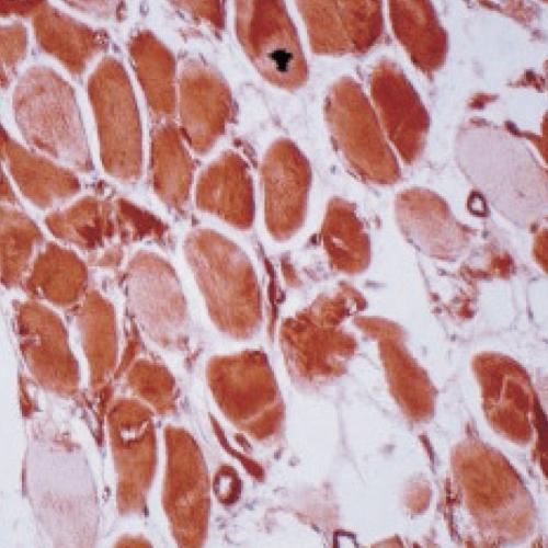
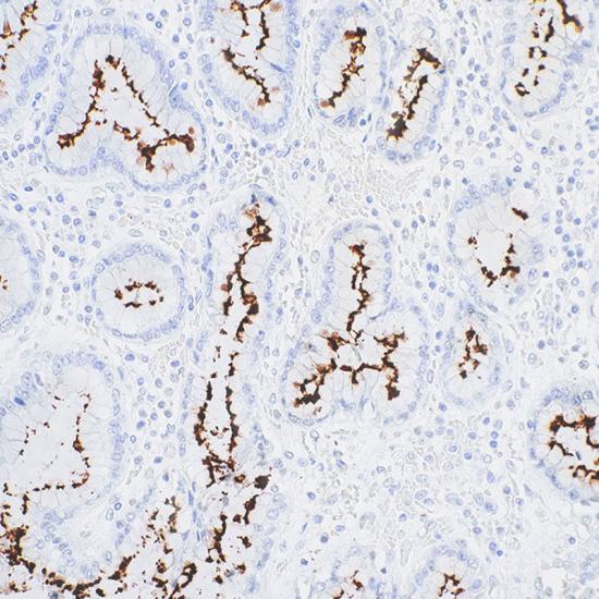
For Research use only. Helicobacter Pylori Antibody reagent is intended for use to qualitatively identify Helicobacter Pylori by microscopy in sections of FFPE tissue using immunohistochemical detection system.
【Principle】
Helicobacter Pylori (H. Pylori) is an endemic helix-shaped Gram-negative bacterium that infected over half of the world’s population. Infection with H. Pylori is responsible for the majority of duodenal and gastric ulcers, and have been associated with increased risk of developing mucosa-associate lymphoid tissue (MALT) lymphoma, atrophic gastritis and gastric cancer. Antibody to H. pylori is useful for detecting the bacterial infection in gastric and duodenal epithelial cells. Add the primary antibody to bind the antigen on tissue sections, and then use HRP labeled secondary antibody binding primary antibody to form the secondary antibody-primary antibody-antigen complex. When DAB chromogenic solution is added, HRP reacts with enzyme substrate to produce brown insoluble reaction product, which indirectly indicating the existence of antigen.
【Main components】
Immunoglobulin, antibody diluent
【Storage】
Store at 2~8℃ for 18 months。
【Sample requirements】
FFPE tissues are usually cut into sections as thin as 3~5μm with a microtome. These sections are then mounted onto glass slides that are coated with a tissue adhesive.
【Protocol】
1. Sample preparation:Deparaffinize the slides in xylene Ⅰ, Ⅱ, Ⅲ for 5 minutes;Transfer the slides once through 100%, 100%, 95%, 75% alcohols for 2 minutes respectively. Rinse slides with deionized water for 30 seconds.
2. Blocking:Block endogenous peroxidase activity by incubating sections in 3% H2O2 solution at room temperature for 5 minutes to block endogenous peroxidase activity. Rinse the slides with deionized water for 30 seconds.
3. Antigen retrieval:Heat the EDTA Antigen retrieval buffer to 100℃. Then place the slides in the boiled buffer and continue to heat for 15~20 min. Naturally cool down for 30 minutes. Rinse the sample with wash buffer.
4. Primary antibody incubation: Drain the slides. Add primary antibody to tissue, incubate at room temperature for 30 minutes. (use antibody diluent or PBS as control). Wash the slides in PBST for 2 times, 5 minutes for each time. If the Primary antibody is concentrated, please dilute it to RTU(ready to use) according to the information on packing.
5. Secondary antibody: Drain the slides. Add secondary antibody to tissue and incubate at room temperature for 20 minutes. Wash the slides in PBST for 2 times, 5 minutes for each time.
6. DAB :スライドの水気を切ります。DAB を 組織に追加し、室温で 5 分間インキュベートします。スライドを脱イオン水ですすいでください。
7. ヘマトキシリン染色:スライドを水切りします。ヘマトキシリンを組織に加え、室温で 5 分間インキュベートします。スライドを水ですすいでください。区別には酸性溶液を使用します。スライドを水ですすいでください。
8.脱水:スライドを75% 、 95% 、 100% アルコール中で2分間脱水します。 スライドを乾燥させます。封入剤を使用して、染色された組織をカバースリップで覆います。
【確実な定位】
1. 陽性局在化: ピロリ菌。
2. ポジティブコントロール: ピロリ菌に感染した組織。
【注意事項】
1. 使用前に説明書をよく読み、キットのすべてのコンポーネントをよく理解してください。操作中は指示に厳密に従ってください。
2. キットまたはキットのコンポーネントを使用した後には使用しないでください。
3. 訓練を受けた専門家のみがこのキットを使用できます。試薬を取り扱う際は、適切な白衣と使い捨て手袋を着用してください。
4. 皮膚、目、粘膜への化学物質の接触を避けてください。
5. 口でピペッティングしないでください。
6. 未使用の試薬、使用済みキット、廃棄物は、地域の規制に従って廃棄する必要があります。
【メーカー】_
会社名: アモイ タレント バイオメディカル テクノロジー株式会社
住所: 36100 中国、厦門市、海滄区、バイオメディカルパーク、2068 Wengjiao Road West、ビル B10、3 階と 4階
電話: +86 592 6315755
電子メール: tlsw@talentbimedical.com
ウェブサイト: www.talentbimedical.com
【記号】
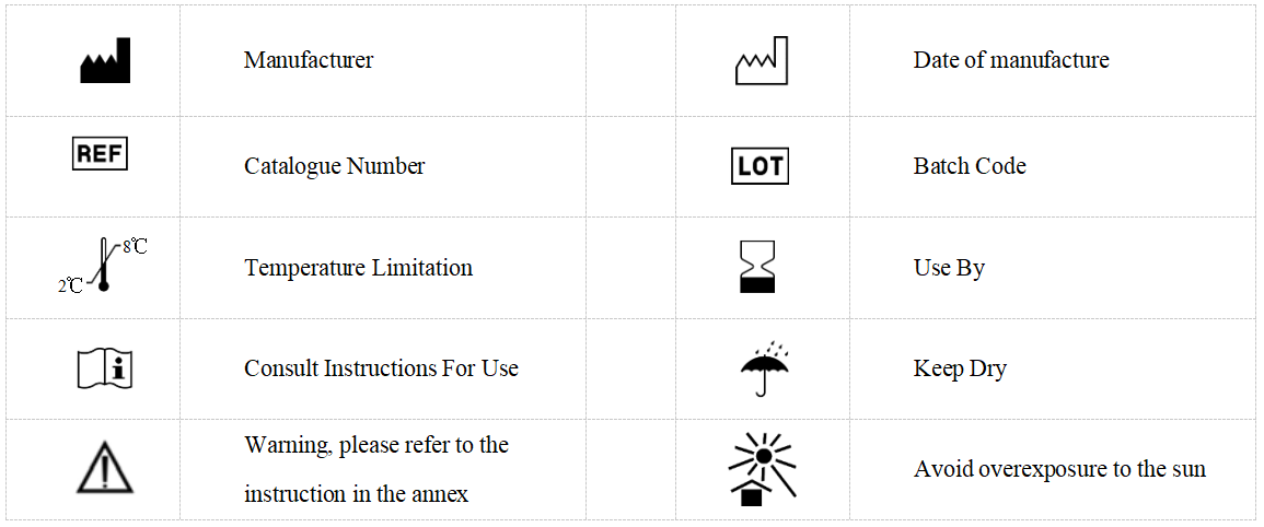
関連するタグ :